Ligand Sequestration Studies in Myostatin/Activin Signaling Models
Animal and cellular studies have used follistatin-based approaches (including protein, mRNA, and gene-expression systems) to evaluate the consequences of myostatin/activin pathway inhibition on molecular markers, tissue morphology, and functional readouts in controlled experimental designs[1], [3], [4], [5].
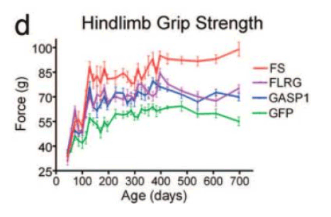 Strength-related readouts reported in follistatin-associated experimental paradigms.
Source:
Oncology Model Context (Expression and Mechanistic Associations)
Preclinical investigations and molecular profiling studies have examined follistatin expression in tumor-associated contexts and evaluated mechanistic associations with activin-related signaling, migration, and metastatic behavior in defined experimental systems, including mouse models[6], [7], [8], [9].
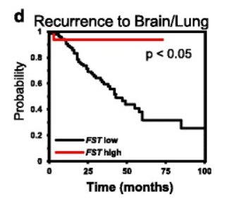 Recurrence-associated probability curves in breast cancer datasets stratified by follistatin (FST) expression.
Cell Proliferation, Fibrosis Markers, and Liver Model Systems
Mechanistic research in hepatocyte and hepatic stellate cell biology has examined the activin–follistatin axis in relation to proliferation-associated behavior, apoptosis markers, and fibrosis-related endpoints in controlled animal and cell-based models.
Developmental Biology and BMP/TGF-β Signaling Context
Developmental model studies have evaluated TGF-β and BMP signaling dynamics in processes such as optic fissure fusion, providing context for investigating how pathway antagonism and related modulators influence morphogenesis-associated signaling outcomes in model systems
Metabolic and Endocrine Pathway Investigations in Animal Models
Preclinical studies have evaluated forced expression of follistatin in defined animal model systems to assess pathway-regulated molecular and physiological endpoints, including pancreatic β-cell biology and glycemic measurements, under controlled experimental conditions
Note: Citations may include a mixture of preclinical reports and broader pathway reviews. Any cited clinical material is provided solely as bibliographic context and should not be interpreted as suggesting clinical, diagnostic, or therapeutic use.